While the first two days were full of departments in every nook and cranny, these last two days were much more mellow, as it took place over the weekend. We started off on a much calmer note, immediately heading towards the dialysis lab, which was one of the few nephrology departments in the entire building, the essential study of treating and diagnosing kidney diseases. When kidneys lose their ability to be able to filter blood effectively, patients are typically referred to the dialysis unit. This certain facility uses machines that artificially filter the waste, toxins, and excess fluids in the blood, basically as a technical replacement for the job of healthy kidneys. This dialysis lab is actually one of the most recently formed departments of the entire department, forming just shy of two decades ago.
The units were equipped with numerous ‘Braun Dialog’ kidney machines, which are tailored for both acute (temporary) and chronic (permanent) kidney failures. We were also taught their specialized procedures, such as how they treated kidney biopsies, and their use of ‘A.V Fistulas,’ which are used to help with long term dialysis. This department was quite large, spanning over two entire rooms that we were allowed to explore. The doctors were extremely nice, and even showed us their prosthetic props for kidneys, which were extremely detailed and realistic, with several parts of it labelled to ensure precision. After we were done meddling with the kidneys, we decided to head towards the reception, the section that was actually second on the list, but was one of the last few departments we went to visit.
We had to wait a lot of time considering that this was sort of a Rush Hour, as there were patients coming up to every receptionist’s stall. After they were done, one of the receptionists directed us to come inside their respective area, and greeted us to the other receptionists. We were allowed to see the sheets they used and how they used quite an old-fashioned telephones to contact doctors, and just like my observership in Kenya a little over a year ago, there were several websites they used to verify insurance policies and their checklist for all the patients. Suddenly a patient arrived and we were able to stand behind the receptionist and see how they treated the patient, directed him and verified the insurance for later patients needing to order medication.
This was one of the longest departments that we went through, but it was so intriguing the way they dealt with patients suffering with grief, with patients who were impatient and angry and how they adapted to every situation. From this experience, I came to the conclusion that the skills receptionists have attained should be applied into every medicinal field, and any field of work in general, considering the kind of people they have to withstand every day. They took us along their personal break rooms and even showed us their interns who we were introduced to. Adding on to that, they all made sure that we all were familiar with all the several systems that they used.
After the receptionist finished explaining everything to us, we moved on to the next department, one of your last ‘long’ departments that we went to, the CATH lab (cardiac catheterization laboratory). The CATH lab was one of the most intriguing and detailed departments, as it is literally the room in which doctors treat heart and blood vessel disorders, with the help of diagnostic imaging equipment, with the primary example of the equipment being the Fluoroscopy unit, which uses continuous X-ray beams to project actual real time moving images of the heart and it’s vessels. While the technician introduced us in, we noticed in the further corner of the room doctors we’re actively performing a surgery, and judging by their tone of voice and the way they looked intently, it was evident this was an extremely delicate operation.
We were told clearly and repeatedly by the nurse at the front of the lab not to make any noise whatsoever or to take any photos, as it’d cause a disturbance. We also saw numerous screens that helped the medical team view the detailed procedure in real time, known as viewing monitors, and there were also a continuous contrast injector that was used for pumping contrast dyes into the cardiovascular system repeatedly. We were also shown many imaging components, such as the DSA, a software able to digitally remove background structures (like bones) so doctors can clearly separate and view contrast dye that moves through blood vessels.
We also saw more examples, such as the C-arm (a large mobile arm that surrounds the x-ray tube on one end and a digital detector on the other, which allows it to rotate around the patient) or the Flat-Panel Detector/FPD (which is another digital sensor that captures the X-Ray beam and converts into higher resolution images) and finally the X-Ray generator (which is a high power unit that energizes the X-ray tube to penetrate deeper tissue tissue and produce clear visuals of the tinier blood vessels). After we were done with this, we were nearly over with the day, and most of the departments we tried to visit were shut down or unable to visit during that time, so we were lucky enough to finish early.
Day 4 Of My Observership
In all honesty, this day was quite melancholy for me, as since I had spent around fifteen hours in this hospital, I felt quite a connection to my team, and I really had treasured the personal moments we all had together. It was such an enjoyable experience, meeting all the doctors and the entire A Learning Lab team was so helpful. They coordinated us with such precision and overall really improved the experience.
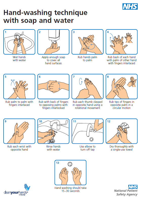
Since we were nearly finished with the entire trip except for the inpatient and another outpatient experience, we determined to finish those as quickly as possible. For the inpatient experience, we went back to the pediatric ward, where they actually let us observe much more closely how they treated and diagnosed patients compared to the last time. They let us into the rooms once the patient left, and gave us thorough lectures on the personal hygiene standards in this hospital, showing us the clean equipment they had to use, their cleaning process with their equipment and their rooms, and their overall sense of hygiene, which was much higher than most hospitals in Dubai.
(Official hand hygiene procedure, proscribed by the National Patient Safety Agency)
They also showed us how several of their nurses who sat at the main desks operated everyday, which was in like routine as the receptionists. They retaught us bedside manner, and their overall journey into nursing services, something just as profound as any doctor, especially considering as how they are much more proficient in medicine compared to doctors after medical school. After we were finished with the entire hygiene procedure and the nurses introductions, we were able to actually interact with the patients and ask them how they were right now, yet not discuss sensitive information such as their reason for visit.
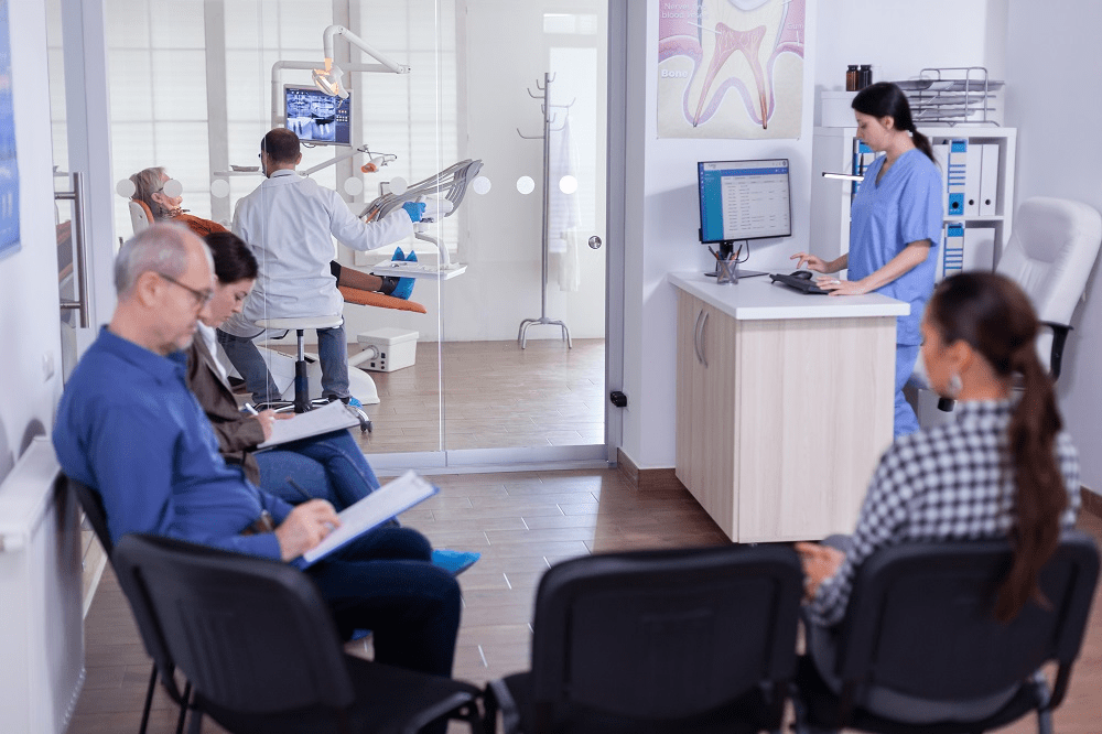
After we were finished with our interactions, we decided to visit (for our outpatient department) physiotherapy, both departments we visited quite early on on our trip. We introduced ourselves to a different doctor, as the previous one had a shift change. After explaining our predicament, he gave us the same tour as the last one, yet showed us through the receptionist area, giving us more explanation and an introduction to how the receptionist directed patients, gave them medication or directed them to different departments if needed to. We saw their hygiene protocol just like we did in pediatrics, and they showed us with much more detailed explanations the machines they used, and we were able to see the more reclusive private rooms, in which we were able to see patients actually working out and saw the posters on their walls of their health regimes and schedules. We were not really able to interact with them compared to our inpatient department, yet they did allow us to observe consenting patients around and actually test out the machines and even view the sauna.
(Typical 4-7 person infrared medical sauna)
After this was done, we had just shy of two hours left, so we decided to go around and actually meet several of the doctors of other departments that we were not assigned to visit. We went to the psychology department and asked around for their brochures, yet all of the psychologists were all fully booked. We then decided to talk to Dr. Bushra, the head doctor who orientated us. She was quite surprised at our efficiency and the fact that we completed all of our tasks much quicker than any of the other groups. We also talked to her about the other amenities A-Learning Lab provided, from tutoring sessions to career advice. After over an hour and half of discussing all this with her, our time was nearly over. In a massive ruckus, all of us handed in our sheets and sent copies of our Emirates ID’s for verification. Then, we all head back home.
It was honestly such a wonderful experience, and my highlight of the summer. Meeting new people and connecting with them on such a personal level in such a short amount of time was something wonderful that happened to me, and every doctor in that hospital was overly nice and welcoming. I would absolutely love to do this next year, and this an experience I highly recommend.
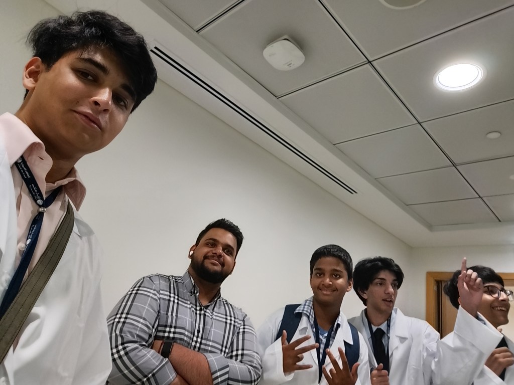
When I arrived in the hospital, I was quite dazed and particularly confused. Most of the staff actually had no clue I signed up for the observership, let alone the fact that there was an observership whatsoever. Yet after going through several blocks and fluctuating through numerous floors, I finally reached the office of the doctor who was actually instructing us, yet at that moment he was completing his regular rounds, leading me and other children (obviously also participating in this observership) to be in a waiting room for around half an hour. These other children were all slightly older than me, which to be perfectly frank made me quite nervous and filled with dread. After all, this was an expensive course, and with being surrounded by older peers, it made me quite anxious, with my heart racing against itself. I’ve never felt like this, even when completing the observership in Kenya, as at least over there I completed my tasks by myself, with no one to embarrass myself against. Finally when the doctor arrived, we all stood in a room feeling quite claustrophobic, where he gave us a brief orientation about the entire hospital and the departments aware of our observership and what we were essentially doing. After he finished speaking, we were all handed a sheet with all the departments and the signature we were required to obtain after viewing each department. We then assorted ourselves into groups of four, (and took a photo with the technician assisting the doctor) and we head on towards the department nearest to us; Radiology, as we also weren’t required to go by the order on the sheet.
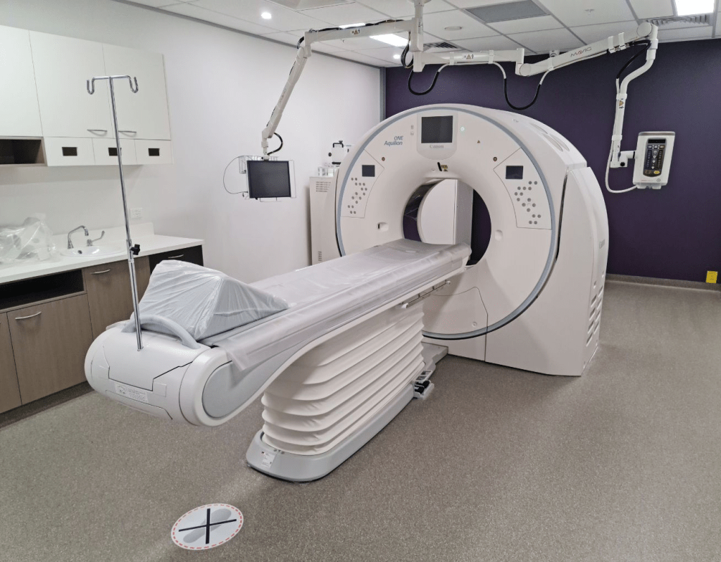
The technician who let us in ushered us into his room, where there were numerous safety warning and several graphs and charts on the walls regarding the dangers of radiology, and after showing us quite a few scans on his computer of the most recent patients and what they came in for, he allowed us inside the CT Scan room, where we saw the actual machine (a large overhead circular shape with a bed like shape attached next to it for the actual patient.)
The CT Scan machine
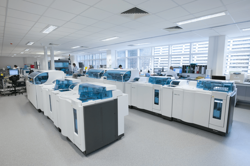
We learnt how pregnant ladies are forbidden to be using it, considering how damaging radiological waves are to a baby. After learning the purpose of the machine, we left this room to be escorted towards the room with the X-Ray machine, where we weren’t really allowed to explore past the doorway. Abruptly, a patient arrived, likely an emergency case, which now led us to be escorted out, without even leaving us any time to actually view the MRI room, yet we just had time to get our signature. Opposite the radiology room was actually the Neonatal Intensive Care Unit, where the nurse took some time after our initial knock. She opened the door, and since another group was likely here before us, she immediately told us to observe in another room. This was a completely empty room, where there was no patients or doctors or any sort of staff. Yet, the wall of this room that connected it to the other room was actually connected due to a translucent black screen, where we were actually able to see an extremely tiny baby, around the size of a SamSung tablet. It was quite still in complete silence, slightly shuffling around the enclosed cot it was inside. After quietly viewing the baby for a few minutes, it started to wail and fuss and turn all around the cot, which led to several nurses having to calm it down and feed the baby. After observing this process, a doctor entered the room me and my team were in and signed our signatures and then proceeded to tell us to leave the room. It was such a short visit yet it was so crucial to me, as the main reason I got interested in medicine was primarily due to my baby brother being sick and meeting the nurses and staff that treated him, and how they worked in such a perfectly synchronized fashion (just as how these nurses were doing a few moments prior.). Right next to the Neonatal Intensive Care Unit, was the actual ICU, which was surprisingly empty when we entered inside. Inside most of the rooms there were cleaning staff who were setting up the television, beds, cupboards and all other necessities but when we actually entered the few vacant rooms there was almost little to no medication stocked inside it and the buttons that are used to call the nurses didn’t work when we tested it. Evidently confused and dazed at why it was so irregular, we asked around the department, who all directed us to the initial nurse who greeted us. She explained to us that the main ICU on a different floor did not have enough rooms to sustain most patients, leading them to form a new one that was still getting set up day by day. Seeing as how we still had quite a lot of time due to this visit and the previous one being tremendously short, we decided to head for the Emergency Room three floors below us. Entering that room made all of us feel quite claustrophobic, as it was quite cramped inside. The receptionist over there was actually quite reluctant to let us inside, yet after we showed proof of the obsevership he let us in, and we walked down the corridor. I noticed Dubai police in the corner of my eye, watching an unconscious person, obviously a reported accident. Luckily before we made our way through the curtains surrounding the vacant rooms, one of the members of my group reminded that even spotting through the cracks of the curtains were technically an invasion of privacy, and considering that literal police were here we decided to quickly get it signed and leave. Thinking that that the ER was going to be our last place yet turned out not to be, we decided to head towards the pathology lab. Seeing as how most of the previous departments were relatively free, this deparment really wasn’t. There were an abundance of staff in every nook and cranny of the ward, and it took us a few minutes of knocking to even be noticed over the constant barrage of voices overshadowing each other. Once they let us in, a nurse quickly showed our team around the department in a hurried fashion, from the machines that tested the blood to the sheets (physical and digital) that they used, before leaving us with the head pathologist.
The following are the most accurate depiction of the layout of the pathology lab, and the types of machines.
He was relatively experienced as opposed to the other doctors, yet he was all the more fascinating, giving us an in depth analysis of what pathology is, and what he was currently doing, yet this was overshadowed by his room having a stench of urine, an experiment unbeknown to us. He explained about his job, the study of abnormalities (something I have highlighted about in a previous post, ‘Introduction to Pathology’), and was instructing us on the machines in use and his day to day life in side of his clinic. After nearly an hour of discussing and him asking questions about our (as in my team and myself) passions for medicine, he left the clinic, leaving us all to go home.
Day 2
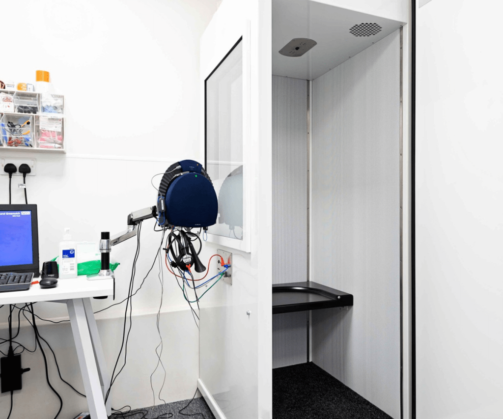
Considering that the first day was relatively complicated due to unknown departments and still getting used to the entire hospital, I expected the second day to be even harder, yet it was thankfully not. After we got registered and instructed on our tasks much faster than we did yesterday, we immediately got started on going to our respective departments. Me and my team, considering that we did most of the grueling work yesterday, thought we could actually meet some doctors privately and discuss more with them, yet since this was a Friday, most doctors (according to nurses or their colleagues in their departments) actually have taken the day off, leading to several departments not having a department head for the day. The closest department for us was the Audiometry one, where we entered first. It was an extremely closeted space, with only one doctor inside who was relatively reluctant to let us into the room. She showed us the room where they test adolescents and adults hearing levels, assisted with a headset, wires and a button to affirm for sounds.
The above is exactly the hearing instruments used and the booths.
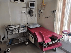
We all took turns testing them, yet the sounds got increasingly milder each time to actually test to what dB level we could hear at. Luckily, we all got similar, great results and she actually showed us pictures of her testing this at a more milder scope with babies in the pediatric and neonatal ward, and even showed us videos of her audio testing on disabled kids in pediatric wards, ranging from the age of (roughly) 6 to 18, which resulted in them obviously hearing less well due to their processing lag. After the brief audiometry session, we were able to quickly enter the Pediatric Intensive Care Unit section, yet it was completely isolated and closed. After calling down Dr. Bushra (the doctor who was in charge of us for that day), she had her own special key to actually open the door and lead us inside that ward and show us around, and it was much larger than the new ICU ward that we had visited the day prior. Several nurses came in while Dr. Bushra was going around the ward, obviously setting the room up, yet there were several machines that did have the nurse button working. Yet, it was still lackluster as it was relatively dark and not as interactive as the other departments. After finished with those two, we nearly had four whole hours left, which led us to start to finish the outpatient department, which was really any department that had patients and if we could observe how they treated them. We decided to head towards physiotherapy, yet that department told us to wait for some time and by the time that we were ready the doctor had to leave after a few minutes within talking to us. We then decided to head up to the internal medicine section, which had surprisingly few people waiting outside the offices. After a few minutes of waiting, we were allowed to visit a free female internal medicine specialist. After brief introductions, she explained her job as a general doctor, and her method of referring, and even showed us through her patients rooms and also introduced us to her entire department. We were allowed to discreetly wait outside rooms sitting in chairs observing how the nurses treated a patient and took them to the doctor. While it obviously had to be discreet to avoid invading the privacy of the patient, we could not really make out what the nurse was saying, and only understood through brief body language. After shifting our chairs a bit closer once the patient was in the doctor’s office, we were able to observe their initial check-up, and then the questions and more detailed diagnosis after learning about the patients new problem. The patients visit was quite quick, with the whole process lasting less than fifteen minutes, yet our questions and answers with the doctor took quite a toll on our whole timeframe, with the whole OutPatient Department took nearly an hour. Considering that it was quite close, we decided to head for the Labour Room, (obviously with no patients), yet this nurse wasn’t informed about the observership, which led to us having show her the doctor’s signature, which took a few minutes before she took us through the relatively cramped labour room, with only one bed, yet around two to three chairs to the bed. The following photo is the closest online reference to the room I was in, as we were prohibited from taking photos in there, considering some lost belongings were still there.
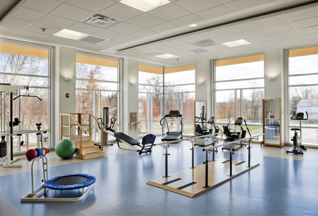
We noticed that there were several machines right next to the bed, which the nurse explained was firstly the heart monitor, obviously to check upon the woman. There was also an IV Pump and several infusion pumps. Right next to the chairs and bed was a tiny separate set up, obviously to keep the baby in after pregnancy. There were also numerous machines for aiding with comfort and mobility; such as birthing balls to help with opening up of the pelvis. The near instant access to staff was also quite noticeable and heartfelt, as there were clear instructions to where to access any member of the department, and a bell for the nurse. She left us to fiddle with the machines and write our notes about the machines and the entire room and it’s atmosphere. While I stated that it was previously cramped, it still felt quite cozy and surreal. After a few minutes, we asked if we could meet any gynecologists, and they actually agreed to that, directing us to Doctor Muhammed, someone who was more than happy to introduce himself to me and my team, and constantly talk about his love for children and passion for gynecology, and a highlight for me was learning he had worked in this hospital just shy of two decades. After we were done conversing with him, we headed on over to the general physiotherapy area, which was a relatively shorter visit than I expected, except for the time we were allocated to the waiting room. After they finally let us in, we were able to actually spot several patient using the aided gym machines (one of the rare instances we actually did see patients inside a department), and the technician working at the moment showed us a full view around the department, from the recovery rooms and other accommodations for the patients, such as a shallow cold pool, and their own makeshift sauna.
Aided Gyms, with several other ‘childlike’ equipment.
Yet, when it came to the largest part of the entire department, the aided gym, he was only able to show us a relatively smaller portion due to several disabled men and women working, majorly on lower body exercises. He even let us into his own personal technician’s office after the tour and showed us old videos of his old patients getting better and their progress from injuries over a few months. He showed the inner bone work that his surgical team had completed on several patients, and he stated himself that his team specialized in lower body (calf, leg, knee) injuries. After this, he directed us to the main intensive care unit, which while obviously busy, and described how several patients transfer from there to physiotherapy. After all this, we were nearly done with the entire day. To end it on a great note, we decided to pursue the nursing department, which was technically everywhere, as we observed a ‘Miss Supuni’ clean and set up several rooms, and mark the attendance sheets. Since she covered several rooms, this was quite time consuming. We actually ended a bit later, as she took us to the head nurse at her section to get the signature.
Pathology, at least when I was first researching professions to write about, seemed quite lackluster as opposed to professions that constantly save lives, it seemed quite boring as it was essentially research. Yet, pathologists especially in the “life saving professions” are sort of how a scriptwriter ties together a movie and they’re usually under appreciated as opposed to the cast and director, and pathologists are also payed relatively lower than other professions. Pathology is the study of diseases, and the cause and function of those diseases and how to cure it. They study diseases using four core step:
a) Etilogy of the disorder, which is the cause of the disease. An example I like to use when describing how the etilogy of disorders are not always straightforward is male infertility. While it can be a genetic disorder, it could also be an accumulation of constant stress and the addition of life endangering substances that they may indulge in, such as alcohol or nicotine. The way pathologists effectively identify the cause of said disease is using their clinical data with more advanced laboratory testing, using a systematic process called clinicopathologic correlation, matching the physical symptoms of their patient’s physical symptoms with their molecular and structural findings. In some cases where the disease is on a molecular, DNA level they use molecular testing, which allows them to identify the forefront fingerprints of the disease.
b) Pathogenesis of the disorder: sort of the sequence of events that the human body and cells go through with biological and chemical changes during the first stage of the disease to it’s final stage. A disease usually starts with it’s initial stage, where the initial stage is usually the exposure, where the disease arrives at an opening such as a mouth or a wound, and it sticks towards the cells using it’s own special structures so it sticks, which is not to be dragged away by the body’s natural cleansers (such as tears, excretion, urination). From now on it breaks through the inital barrier and enters the deeper tissues. From now on the disease/pathogen will replicate rapidly and secrete toxins that destroy the tissue directly.
c) Morpholic changes; now these are the changes that due to the disease, pathologists are actually able to see, meaning the physical changes and differences in cells and organs. (These changes are though seen on a microscopic level).
d) Functional derangements; while relatively similar to morpholic changes, they are how the body behaves differently and out of order due to the disease, which are essentially symptoms of the disease. The main symptoms of functional derangements are usually that the patient will naturally feel short of breath, tired and the heart mispumping (extremely irregular for even patients with deadly disorders). Yet on an organ level, if the heart muscle suffers from morphologic change it will directly lead to heart failure (and this sort of change can also occur in other major organs).
Evidently I am not as educated on pathology as I am on a subject as neuroscience or even cardiology, yet it is truly fascinating and the backbone for all medicine.
As anyone can tell, I am an avid fan of neuroscience yet I have never really spoken about neurosurgery which is just or even more fascinating. What better way to start discussing topics about neurosurgery than discussing emergency issues, something I also enjoy discussing.
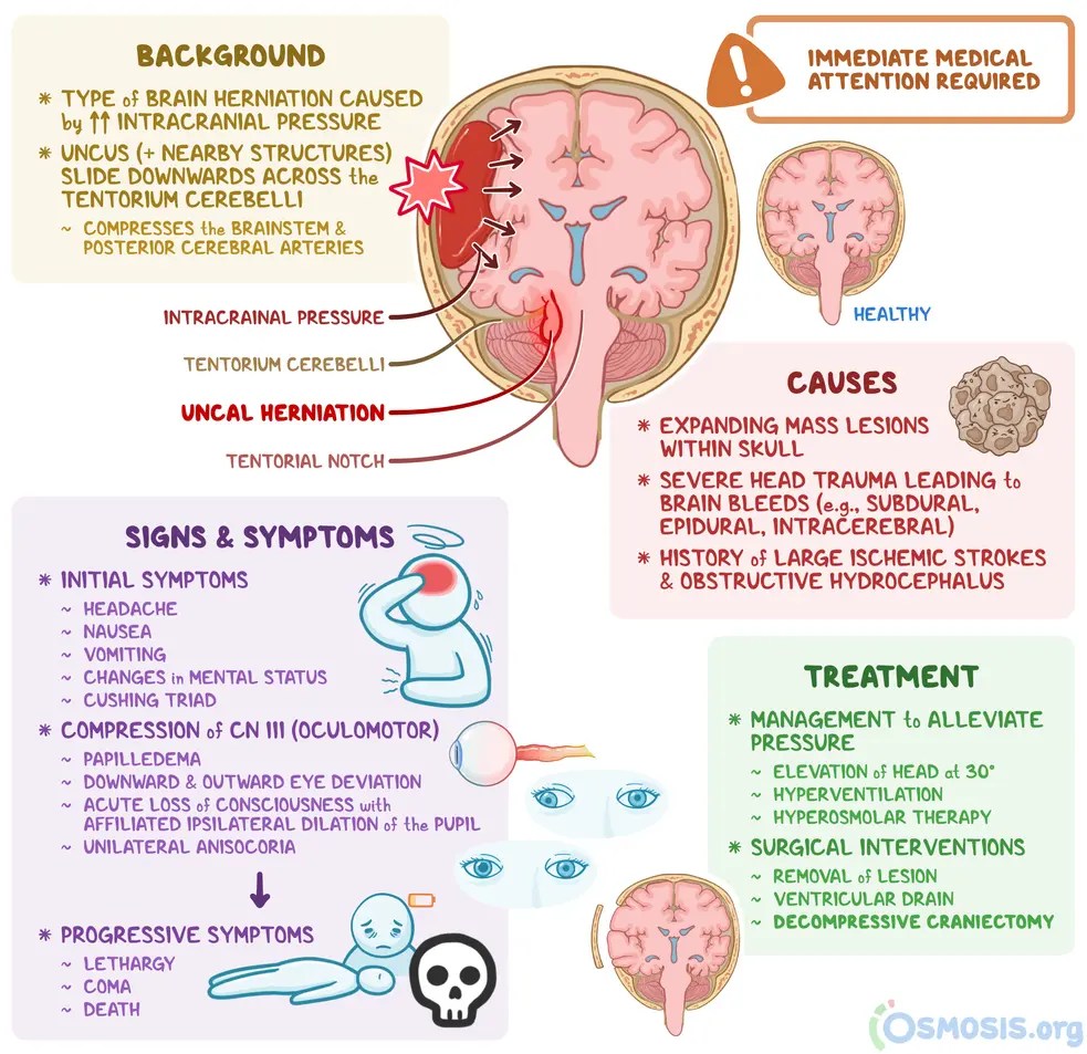
When understanding head trauma, we must first talk about the rigidness and the detailed biology about the adult and even the adolescent skull. Unlike an evidently expanding abdomen (which would be soft) a cranium is a fixed vault of solid bone. Inside the cranium there are three primary components that share a quite limited volume. Starting off with the brain tissue and intracellular fluid(also known as brain parenchyma), which takes up around eighty percent of the brain, and then the blood and cerebrospinal fluid, which each take up the remaining twenty percent. Another doctrine, known as the Monro-Kellie doctrine started that since the skull cannot expand the total volume inside should remain constant. Yet, traumatic impact is still able to introduce a fourth element, usually a swelling brain or an unusually rapid growing blood clot (known as the epidural/subdural hematoma) another substance in the brain has to yield to break through. Adding on to this, when a brain injury does occur the body immediately attempts to adapt to this added mass through two main spatial compensation mechanisms. The primary being CSF displacement, where excess cerebrospinal fluid is squeezed out of the cerebral ventricles and down into the spinal tecah. The latter is venous compression, where low pressure cerebral veins and dural sinuses are compressed, which forced blood out of the skull into the internal veins. During this immediate stage, patients actually remain weirdly stable. Continuing, as the bleeding and swelling the spinal fluid and blood shift away, and then intracranial pressure remains. Yet the high pressure inside the skull is not just uncomfortable. This pressure completely limits the brain cells of oxygen. Brain tissue relied entirely on CPP(cerebral perfusion pressure) to deliver oxygenated blood and glucose across capillary beds. CPP means the pressure gradient that drives blood flow through the brain, calculated in the following equation.
CPP=MAP-ICP
ICP is intracranial pressure, which when disrupted compression tissue and vital microvasculature. MAP on the other hand stands for the mean arterial pressure which when disrupted completely drops during systemic shock which reduces pushing force towards the brain. CPP when disrupted causes oxygen starvation, clinically known as cerebral ischemia.
Now, neurotrauma clinicians divide traumatic brain injury into two phases, primary and secondary. The primary phase is whenever the immediate damage is caused by physical impact and happens in milliseconds. This cannot be reversed yet it can be repaired. Secondary injury is when destructive cellular mechanisms that unfold over hours and days following the impact.
There are also two types of brain swelling, also known as cerebral edema due to low CPP. The first is Cytotoxic edema, which is when sodium rushes into the brain cells to pull water in through osmosis and causing individual neurons to swell due to lack of oxygen disabling the cells ATP dependent membrane pumps. The second type id when inflation breaks down the blood brain barrier. This means plasma proteins leak out of damaged white blood vessels into the space which draws fluid into the surrounding white matter. As the swelling progresses, any unchecked pressure can cause brain tissue to be forced through internal bony boundaries which is also clinically known as brain herniation.
Now coming back to the clinical treatment side, managing acute terminal brain infections is essentially surpressing ICP levels while keep CPP above it’s optimum amount. The main interventions medicals team use are the following;
I seriously believe traumatic brain injury is truly a highlight on how closely physics biology and surgical medicine can all closely come together and work off each other, and the precision and detail into this subtopic is really breathtaking, with several layers of depth I would likely never be able to cover. Truly wonderful.
As I have stated in previous blog posts, the human brain during childhood is extremely sensitive and vulnerable, meaning “regular” habits picked up as a child can never be broken even as an adult. This does also mean that the good habits can be picked up, and the brain adapts so much easier as a child as opposed to an adult or even a teenager (this is called neuroplasticity).Yet, this also means that the brain’s baseline electrical environment so much easier to be broken down, and when that balance breaks a seizure occurs. In general, in a healthy brain, every thought and movement and emotion occurs onthe balance of two chemical signals, the glutamate and the GABA, with the first being the one that pushes neurons to fire and pass meesages along wnd the latter being the one that calms neuronal activity and prevents overload. In a brain that undergoes seizure activity, mild disruptions in ion channels that control the flow of sodium and calcium cause the brakes to fail, which causes the glutamate to get stuck and neurons to fire simultaneously in a quite high voltage electrical chain reaction.
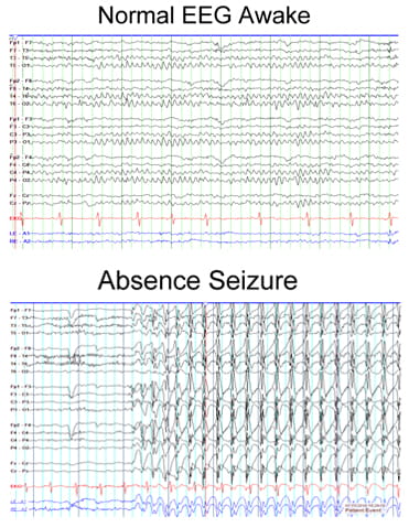
Evidently, to make sense of this neurologists use an electroencephalogram (EEG), that can translate complex brain waves into physical patterns on a hospital screen by placing small sensors across the scalp that detects the electrical signal. When seizures occurs the waves are able to identify and differentiate them.
The first type of seizures are called the Absence Seizures, where the wave pattern is a 3 hertz spike and wave, where a sudden synchronized loop is bouncing between the brain’s core and surface. The next main type of seizures are called focal seizures, which are often tied to a structural site, with their wave pattern usually being focal sharp waves, where a localized electrical spark originated from à precise spot. The last type of seizure I will be talking about is the type of infantile spams, which require immediate intervention. They are completely unpredictable and are electrical chaos. By tracking where the signals actuwlly start, doctors can isolate the seizure onsent zone, which pinpoints whether a seizure is localizing to a single region or firing across both hemispheres at once.
Back to a point in the first paragraph, neuroplasticity. While this can be quite a significant challenge to a child, it can also be an asset. If electrical misfires happen over and over, the brain can accidentally learn this habit, and this concept means that untreated seizure pathways can carve deeper holes into the brains wiring over time. This, on a positive note, also means that à child’s brain can re route it’s connections around damaged areas, which means they can recover functions an adult brain would lose forever.
Yet, mapping these patterns isn’t just academic, it helps dictate how we treat the patient. Most doctors start with targeted medications, as certain drugs calm specific ion channels, yet choosing the right one depends whether the seizure is focal or generalized. Some doctors also enforce metabolic shifts, meaning a specialized diet that targets GABA levels. Now if the seizure origin is locked to a single spot, modern neurosurgery can remove the target zone entirely.
As you can tell, neuroscience to me is the most fascinating field in medicine, and epilepsy is such an intricate and complex disease, with childhood epilepsy even more so. Learning about this is special to me, as my own brother had been diagnosed with epilepsy in December of last year. Learning how to read the cellular dynamics and electrical patterns on EEG waves, I’m glad doctors move closer to protecting the mind when it’s most resilient, yet vulnerable.
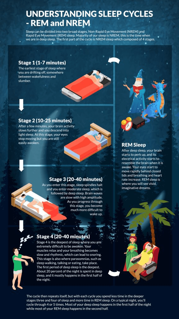
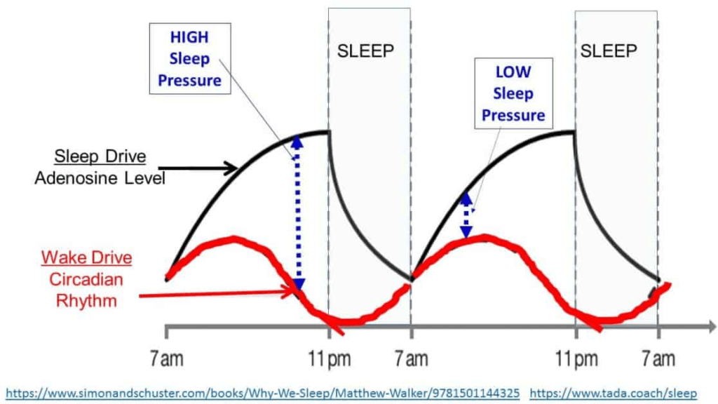
Sleeping is not a mechanical, ordinary cycle that can easily occur at any time, it’s a chemically structured and synchronized system. Hormones, such as dopamine, seratonin and acetylcholine repeatedly rise and fall throughout the night to shape each stage of sleep, with an example being REM (deep sleep), which acetylcholine is high of. That’s why many sleep specialists recommend not using screens before bed as it can disrupt the brain’s dopamine regulation and dilute the mind’s circadian rhythm (perception of whether it’s daytime or nightime). There are 4 cycles of sleep, the first three phases of NREM sleep (non rapid eye movement, and is when the brain’s glymphatic system starts going to work, and even the cerebrospinal fluids are able to flush out metabolic wastes, after being triggered by a drop in noradrenaline. The waste clearance rate during NREM sleep is roughly about 60% more effective and apparent now as opposed to when awake) and then REM sleep, which is when dreaming occurs. During this time, the levels of monoamines such as seretonin drop whereas acetylcholine levels rise much higher. Memory integration, the most important part of sleep occurs during this time, and research also shows that targeting dreams during REM sleep usually boosts creativity and problem solving. NREM sleep uses slow oscillations for its purposes whereas REM sleeps uses theta rhythms to help with their integration.
Speaking of how sleep helps with memory integration, the main reason people forget their actual dreams are due to two main reasons. The first is that the brain usually has their dreams in REM sleep as opposed to NREM, and they switch in cycles, meaning that when someone actually wakes up their REM sleep cycle had last been switched long ago, and unless the person suddenly wakes up in the middle of a REM cycle, they will likely forget their dreams a few second into waking up. The second reason is that the brain forgets most things that we deem as irrelevant, and those dreams being quite unrealistic are forgotten often just due to the brain not wanting that to be confused with real life.
The main transition and switch in the brain during sleep is actually how we are able to transfer between being asleep and being awake, which is mainly due to the actions of the ‘flip-flop switch’. This ‘switch’ relies on two different groups of neurons that silence each other mutually. On one side , the neurons are the ‘awake’ monoamingeric nuclei(such as the locus coeruleus, dorsal raphe, and tuberomammillary nucleus) and on the other side are the sleep promoting ventrolateral peroptic nucleus. Due to the fact that they actively inhibit one another in a synchronized fashion, it creates a sort of switch mechanism.
This sort of mutual inhibition makes sure you are either fully asleep or fully awake, meaning there is no such thing as ‘half-asleep’ no matter what movies may suggest. Now to keep this fact true, the oxerin and hypocretin neurons that are located in the lateral hypothalamus act as stabiliziers. They effectively reinforce the wake side of the flip flop switch when you are supposed to be awake, and the same applies for the asleep side of the switch. Adding on to this, while the switch does handle the rapid shifting between awake and asleep, it’s triggered by adenosine, which builds up your natural tiredness throughout the day until its time for you to fall asleep.
In conclusion, the neurological basic and sciences behind dreams and sleep in general is really fascinating, highlighting to me the fact that while our body is complex enough while awake, there are several other factors that come into play while we are asleep which are equally as intriguing.
In all honesty, Neurology was not a field I had so much knowledge in compared to other fields, such as pediatrics or even physiotherapy. Sure, I had participated in that epilepsy awareness event, but my knowledge was just minimal. I mean, sure I was relatively familiar with mental disorders and whatnot, but having enough knowledge to write a whole research paper? Yeah right….Well, it’s been over three months since the awareness event, and I am glad to say what I had previously thought near-impossible, I have relatively completed.
The thing is, Neurology is such a complex subject with so many sub-branches and other topics, it’s near impossible to know every single thing about the brain, yet alone contain all of it into a single research paper. The brain is such a diverse area to study, so the main subjects I wanted to put into this paper are
Disorders
Aging
Social/Emotional Aspects of The Brain
But to state something before I start; I am not the Brad Pitt of neuroscience whatsoever. Even all this knowledge I am boasting about is quite minimal to what actual neurologists have to learn to actually become a neurologist. Yet, neurology is such a fascinating subject, there is always more to learn as the brain evolves and grows. Without futher ado, let’s start this.
Mental Disorders
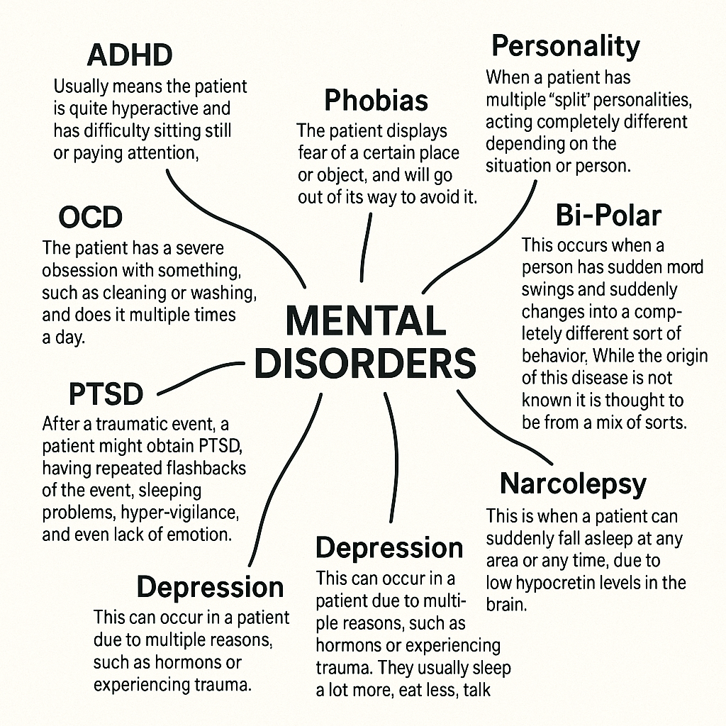
In the brain, there are certain parts of it assigned to certain areas of our life, such as thought, speech or comprehension. When these parts get damaged, whether that be through self-infliction or car accidents, they can lead to brain damage or impairment, which can lead to disorders. Most mental disorders are relatively harmless, such as attention-deficit-hyperactive-disorder (ADHD), not being able to sit still while watching a documentary, or obsessive-compulsive-disorder (OCD), having an obsession with cleaning your glass-model cars. Yet, some can end lives or indirectly cause massive bouts of harmsl such as schizophrenia, alzeihmers, or shock paralysis (yes, it sounds exactly like it’s spelled.)
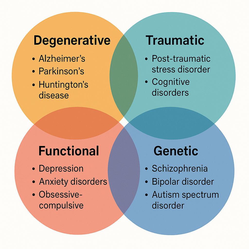
Yet, there are more reasons for disorders, and the most common of them are
Degenerative; These are mostly in older people. They are due to inactivity of their brain or muscles, making them more prone to crippling and making them weaker as a whole. These mental malfunctions can unfortunately accompany them, such as Alzheimer’s, tending to forget things as mild as grocery shopping to as severe as their own name. While contradicting my previous statement, not all of these disorders are technically ‘deadly’, yet they increase the risk of the elderly person too commit dangerous acts such as self-harm or reckless driving. These diseases are mostly present in elderly people over the age of 70, yet it’s not exactly uncommon for them to be within the range of 50-69.
Genetic; While the most common mental disorders are due to other factors, there are multiple disorders that can be contracted due to genes and genetics. These disorders are mostly uncontractable, yet there are other ways that they can be contracted other than genetics. Yet, just due to the fact that one family member had the disease does not mean that you will surely get it, as it can just be a one-time thing. These genes are mostly common in men (86%), yet some women also contract it.
Traumatic; These sort of disorders are quite common, and caused by experiencing traumatic events. These events can range from car accidents to war-time. Now, just experiencing these events does not directly mean that the patient will contract this disorder, yet it’s quite common to contract these disorders after these experiences. The most common disorder out of this type is post-traumatic-stress-disorder (PTSD), which have the symptoms of habing repeated lucid dreams about the events, and not being able to function properly, and mostly only being able to speak about the event.
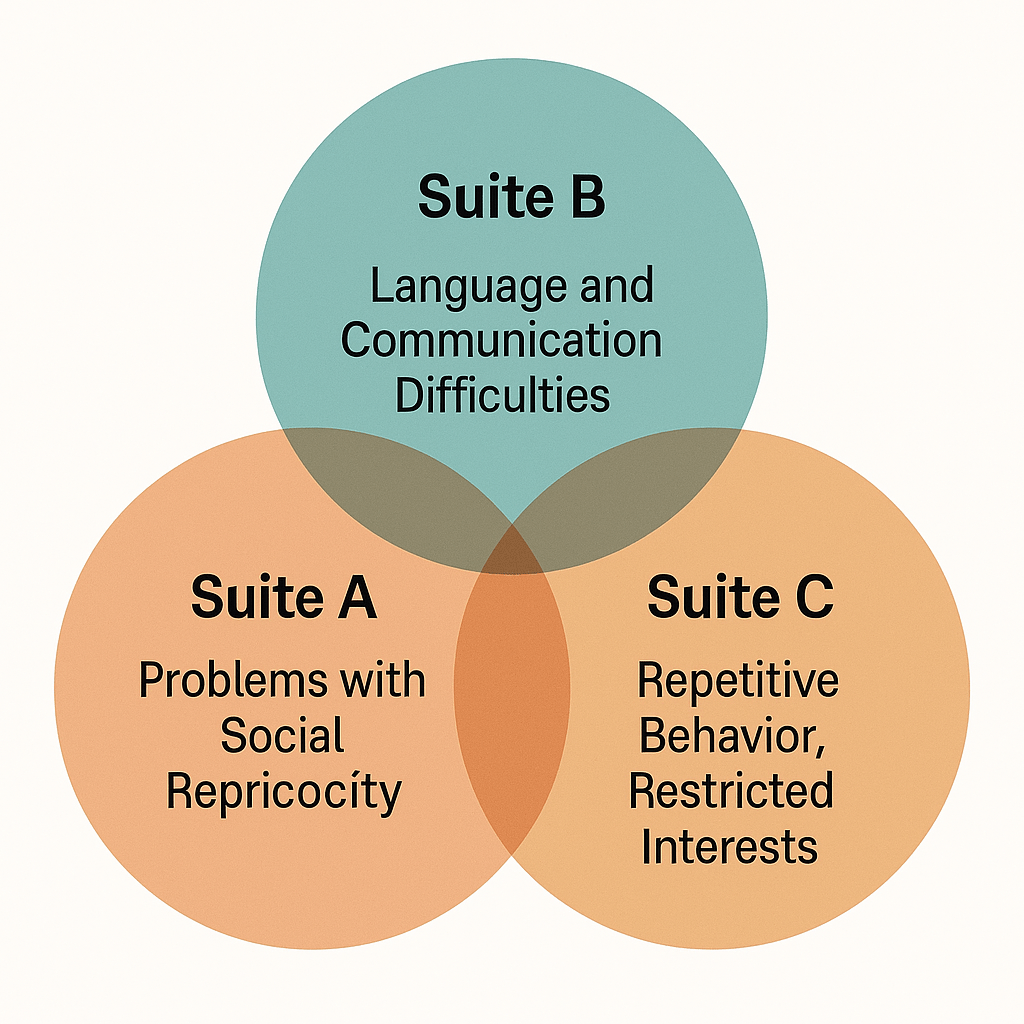
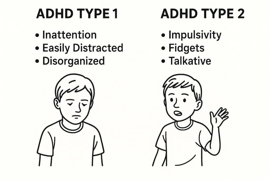
Now, there are multiple disorders that can be diagnosed on a spectrum, noticabely autism. Some people without autism show different forms/suites of behaviour, and the common behaviour in all suites lead to being diagnosed with autism. The common suites are presented in the diagram below.
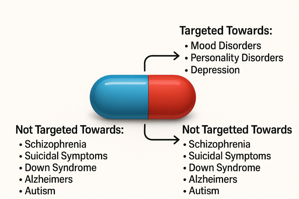
Evidently, there are treatments to these diseases, yet it’s not proven they have a perfect success rate. They mostly have quite a number of side effects, commonly being very emotional, tired or feeling very nauseous some time after taking the medication. Yet, this does not mean these meds are never presicribed, it’s quite the opposite. They are prescribed quite often, especially in anxiety disorders, or depression. They are never a permanent cure though, they are always part of a larger treatment plan to help ‘cure’ the disorder.
The most common medication type used are pills. While they are commonly used in the disorders explained above, they can also be used in ADHD or OCD, yet not as often. They help and calm the patients, and help deactivate the depressive hormones and increase dopamine, the hormone that makes you feel optimistic. These medications help regulate brain chemistry and reduce depressing/distressive symptoms in the patients, and overall help them function normally, though these medication obviously will not work for all disorders.
Now, the diagnostic aspects of the brain. Mental disorders as a whole cannot be identified just from a few symptoms; they have to genuinely interrupt everyday activities, such as school or dinner, and contain most symptoms to be properly identified. Usually, some physical tests by doctors are used to identify these disorders, yet multiple certified online tests can be used, though they could be inaccurate, or the patient self-diagnosing themselves. Though, eve after online diagnosation, it is evidently best to consult a local psychatrist or doctor. Adding to the symptoms aspect, not all symptoms have to be present. In ADHD, only a few symptoms are ‘compulsory’, such as inability to stop fidgeting, to be diagnosed with a certain ADHD type.
Yet, with more complicated diseases centered towards the brain sucb as epilepsy or schizophrenia, brain imaging scans are often used, such as CT scans and MRI’s.
In medieval days, mental illnesses are commonly regarded as the disease of the inner spirit during the middle ages. It was assumed that devils, or ‘foul spirits’, would enter a persons body, and make them ‘insane.’ The physical theories of mental illness were;
Imbalance of the four humors (which back then determined a persons health and wellbeing)
Blockage of multiple supernatural, healthy ‘forces’ and spirits, thought to be because the patient was a witch or some sort of demon
It was really in the nineteenth century, when the acclaimed Sigmund Freud thought up the idea of a subconcious. He developed the foundation of psychoanalysis, the idea of bringing hidden ideas to the consciousness, and the building block of therapy nowadays. The most common (and highly ineffective) treatment back then was hypnosis, and it was due to the invention of brain-scanning devices that proper treatment for disorders started to be used.
A reason why diagnosting is so important nowdays is the way it impacts indivuals, such as in acts of law. In severe cases of sexual assualt or murder, these alleged ‘criminals’, can technically have no charges pressed against them is if they are mentally damaged, or pleaded insanity. This can also decide upon multiple other important factors, suchh as whether they are suitable for certain types of employments (would someone with severe alzeihmers be quite suitable for customer service), or even eligible for government (or state aid).
Surpisingly, mental illnesses can also affect aging, especially in younh children, pre-puberty/adolescent. The way it impacts is by making the body phsyically incapable, passing aging checkpoints (i.e first steps, first words, jumping) way after it ideally should have done. Around 34% of autists are shorter than average and with less than 7% being taller than average.
}”
Now, I personally think I am finished with this topic, so it’s about time we move on.
Development and Aging
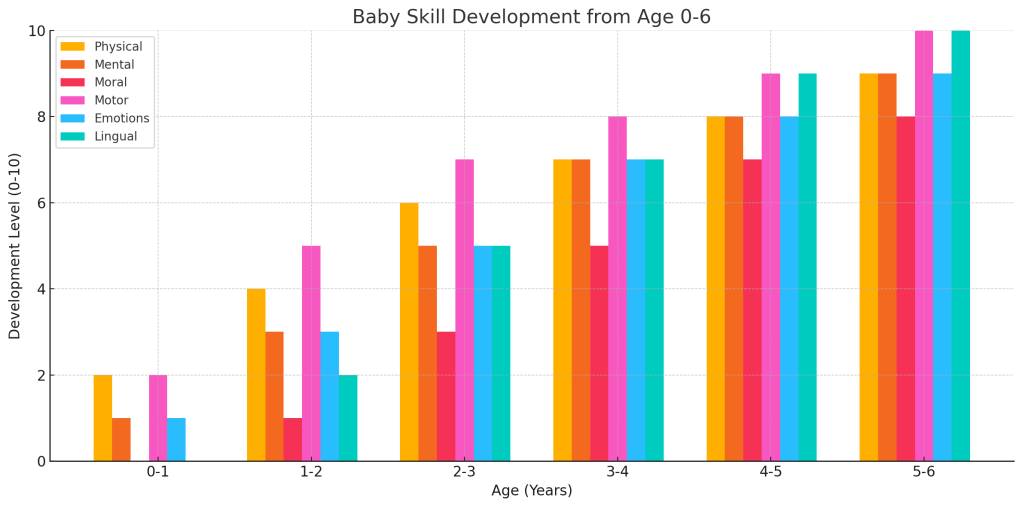
The human brain and body evolves and grows slightly differently deending on the person, but the brain development always stays the same. While the embryo is in the uterus, it slowly starts to develop its brain, starting with the neural tube, and slowly forming other parts such as the brain stem, cerebellum and cerebrum, with the sensory nerves also starting to develop. Finally, during birth, the baby develops nearly all 100 billion neurons, the same as an adult, yet nearly all of them are not mature, and develop until around the age of eighteen. The baby obviously starts to grow, and understand topics, developing multiple of the neurons, maturing quite rapidly. Yet speech, and a lot of similar characteristics are automatically wired into the brain, yet evidently all babies require appropriate simulation to actually develop the characteristics.
Now, the next step of growth is childhood to adolescence, where children develop motor skills and much more concepts that are extremely important during these years.
Yet during childhood (five to eleven years of age), there is not much significant development in the brain, except for noticable skill development and maturing through childhood experience, yet it’s really during the teenage/adolescent years that the body significantly changes like it had done during infantry.
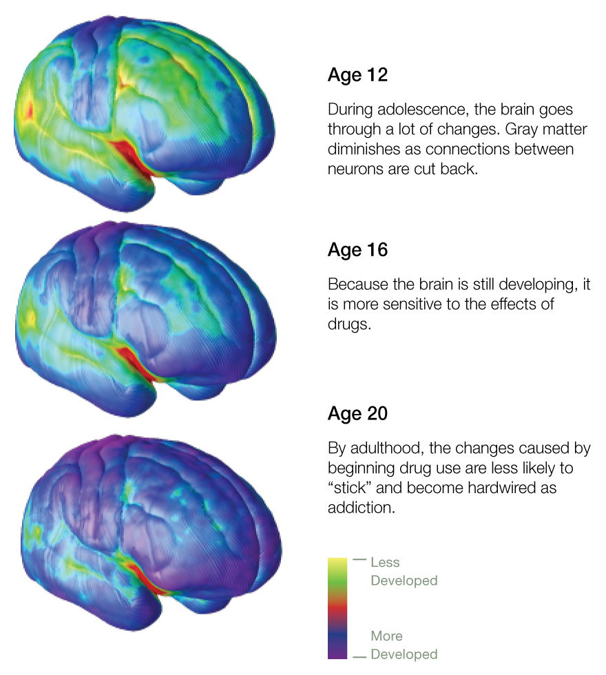
Between puberty and early adulthood, the human being goes through a renaissance-like stage. This process is well reflected in impulsive and rebellious behaviour, and eerily sudden personality changes. While all these changes take place, the teenage brain is quite vulnerable, and personality traits like risk-taking may be amplified to the extent that they can cause serious dysfuncton such as addictive drug-taking, criminal-like behaviour, and intense anxiety or depression. Fortunately, in many cases, the issue passes as the brain matures, yet it can sometimes signal the unofrutante start of long-term mental health problems, that can last even throughout adulthood. These changes in the teenage brain (in both genders) are driven by testosterone release, and the hormones make neural pathways extremely flexibile, in the sense that connections can make or break quite easily. On the positive, this allows teenagers to learn new things quickly, and to adapt to new habits and personality traits. Yet, on the negative side, this can lead to easily being influenced, and being more inclined to risk-taking, rebellious and somewhat pessimistic behaviour, which can lead to self-inflicted trauma, in severe cases.
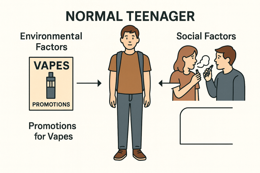
Their pre-frontal cortex is still developing, which is thought to be one reason for teenagers to be more inclined to this sort of behaviour, yet it’s obvious this behaviour can be present due to an addition of enviromental and social factors. The pre-frontal cortex is closely connected to the basal-ganglia which plays a quite important role in the development of motor skills. The fibre that links the two hemispheres in the brain-the corpus collosum-thickens, allowing for increased information to information processing skills.
During the teenage years, the patients have a high risk of being diagnosed with a mental illness, commonly ADHD and Anxiety, yet when teenagers experience traumatic events, they are extremely likely to contract a severe disease such as PTSD or Schizophrenia. When these patients near early adulthood, they are likely to contract a substance addiction, commonly alchohol, drugs, or cigarretes.
Most teenagers (commonly women), tend to get quite emotional very easily, thought to be due to the limbic system in the brain, which helps control our emotions. During puberty, the system is surprisingly active compared to any other time period, and this leads to the emotions being extremely prominent, and outbursts of sudden rage, or sadness occuring quite often, During early adulthood, the system matures, helping handle emotions significantly better.
Now during adulthood, the brain doesn’t stop growing. Like any other organ, the brain continues to reform itself long into adulthood. New brain cells continue to be created, and the architecture of the brain is changed constantly in response to life experiences. Humans brains are, compared to most animals, are quite slow to reach adulthood. The prefrontl cortex is the last part to actually become fully active, and full myelination-the sheathing of neuronal connections, which allows information to flow freely along them-does not occur until late into their twenties or thirties. Once the pre-frontal cortex is fully online, it becomes more active in situations that require emotional contenrl Whereas a teenage, or even a child might be overwhelmed by emotion, the prefrontal cortex inhibits emotion when necessary, and so allows for a more thoughtful, deliberated response.
Now, with the creation of new brain cells allow new information to be stored, but their arrival disrupts exisiting memories form in the hippocampus and are tranferred to long term storage in other brain areas. For a while, the memory resides both in the hippocampus and elsewhere. After a few years, the memory is cleared from the hippocampus. Until the memory is fully transferred, the arrival of new cells in the hippocampus may weaken the connections encoding memories stored there.
It is thought that the number of brain cells in the adult brain was fixed early in life and that laying down new memories and learning new things was achieved entirely by changes to existing neurons and their connections with one another. While this sort of rewiring is important for learning, it is now known that adults also benefit from the creation of new brain cells. Neurogenesis, the name for it, occurs mainly in the denate gyrus of the hippocampus. About a third of the neurons in the adult hippocampus are replaced in a person’s lifetime. Adding to this, a person’s brain continues to mature until their late twenties. The main changes take place inside the higher functional areas of the brain, such as the frontal cortex, which gradually becomes more active-pulling together information from the rest of the brain and forming a complex and holistic view of the world. Until then, the emotional parts of the brain are not fully connected with those areas concerned with thought, judgement, and behavioural inhibition. As the connections between the areas become more stable, people tend to react less emotionally and impulsively-instead becoming more cautious, considered, and exercising better judgement.
Now, onto the ageing brain. See, the traditional normalized view (probably thanks to the completely medical accurate ‘Simpsons’) is that the brain and body start to sort of degenerate. This is relatively true, but in the fact that neurons are ..’lost’, and for those that remain, impulses are transmitted much slower than normal. This obviously leads to slowing thought processes, memory problems, and deteriorating reflexes, which causes problems with physical aspects of the body, such as balance and movement.
In the past (meaning ‘cursive handwriting’ level old), it was quite rare for people to live above the age of 50, so we have not evolved much to use the brain in such advanced years. This makes the ageing brain quite a relatively new phenomenom. The degeneration of the brain and nervous system is not caused by a diseasee, and so should not be confused with the pathology of dementia, (which, in fact, is associated with patterns in specific brain changes). Now, recent research shows that most neurons actually remain healthy until you die, but the heart volume and size decrease around 6-10% from the age of 20-90.
One weird thing is that age can affect excitement levels (not in the way you think of, where old people sit around gossiping and yelling at children riding bicycles), but in a scientific kind of way. Dopamine, the neurotransmitter that triggers excitement, rapid decisions and overall happiness. Brain-imaging studdies show that as people age, their dopamine circuits decrease. This is associated in behavioral changes, as dopamine is associated with thrill-seeking and risk-taking. Neurologists believe it is due to the fact that older people seek a quieter life, or even a less thrill life as their dopamine is less abundant. An example could be any holiday where you recieve presents, such as Christmas. Opening presents is highly exciting for childrens and adolescents, but evidently much less so for adults and the elderly, as dopamine is triggered by rewards, or gifts, which also has much less impact as you age.
Now, ageing is not all pessimistic as I had explained before, and there are also positive aspects. The brain can compensate for the effects of ageing, and mental function can even improve with age. Myelin increases in the temporal and frontal lobes in the 45-50 age group could enable people to manage their knowledge better. Not only that, comprehension studies have shown that high-functioning elder adults use both brain hemispheres, which is sort of the brain’s memory to keep thought and memory processes stronger, an apology for making most of the body quite declining and useless.
Now, even newer research into ageing shows that the rate if decline could be slowed by positive lifestyle factors, such as regular exercise. Research has also found that reducing food intake, resulting in lower blood glucose levels, may slow the pace of change, as blood glucose can cause damage to enhancing proteins. Certainly, people with elevated blood glucose lvels, such as those with T.1 diabetes show more signs of brain ageing than non-diabetic people. There are evidently multiple benefits of a healthy lifestyle, such as simulating neural tissue growth. Gentle aerobic exercise, such as rapid walking, regular sleep, a healthy and balanced diet are expected to help delay age-related mental issues such as memory loss.
Now, while the next few paragraphs of this next topic are not exactly connected to the ageing of brains, but rather the ‘Brain of the Future’. While it sounds extremely cliché, the fantasty that has existed since 1984 with The Terminator is now extremely close to being real. As we discover how the brain works, the prospect of changing it, enhancing it, and developing artificial brains is quickly becoming fact rather than fiction, as stated before. Technologies for mind reading, thought control, and AI machines are already with us, and are becoming more sophiscated every day.
When a person is thinking, their brain produces mild electric signals. Neuroscientists have discovered ways in which the eletric signals are able to be picked up by sensors and sent wirelessly and sent to other electrical devices, making it quite possible to move or alter objects by literally pure thought. Most research in this field is directed towards developing devices that are able to help people with nervous-system injuries regain the use of limbs previously paralysed. The technology has also been picked up by multiple game factories, who have produced games that can be played using thought power.
To add to what explained previously on mind reading, the way it’s made is through the neural activity. The ‘picture’ of neural activity created by fMRI scanning can be sort of translated into a accurate description to what a person is seeing and thinking (to some extent). The way this is formed is that the output of the scan, captured while they are looking at a particular image, is processed by sophiscated computer software that translates the pattern of activity into a sort of ‘read aloud’. Such ”mind reading” is made possible as neurons in the visual cortex are specialized for specific stimulti–horizontal/vertical lines, so their firing patterns are indicative of the tyoe of visual stimuli the neurons are registiring. This also helps with lie detection. Now, finally onto the next topic.
The Social & Emotional Aspects
Emotions are generated in the limbic system, explained before as used a lot in adolescents to generate emotionally type responses. Each emotion is produced by a different ‘network’ of brain modules, including the hypothalamus and pituitary gland; which control the hormones that produce physical reactions such as increased heart rate and muscle contraction.
Now, we have evolved a ‘conscious’ emotional system, yet we still retain the primitive, automatic responses at the heart of emotion. An example could be hearing a frightening sound, we register it into the amygdala before we are even conscious of it. While the sensory information is sent to the cortex to be made conscious, the amyglada sends messages to the hypothalamus, which trigger changes that ready the body for fligh, fight, or appeasement. This quick route allows us to take instant action to save ourselves. When we ‘start’ at a loud sound, then relax on realizing that it is harmless. we are experiencing both stages-conscious and unconscious responses and reactions.
When it comes to social and emotional aspects, emotions in nearly everyone are quite conscious, and we’re usually aware of what emotions we’re feeling at every moment, but they’re not something physical, just our brain’s natural response to stimuli, designed to push us away from danger and towards reward or comfort, hence the term comfort zone. Emotions are generated quite constantly, yet there are so much we’re only aware of the predominant ones. (When I say ‘natural response’, while I do mean what our brain’s naturally designed to, but the brain as said before is also affected by environmental factors. Meaning if someone grew up in an area where danger was quite normal and encouraged, the brain will adapt to it’s surroundings and push us to danger.)
Now in itself, emotions are generated in our limbic system, used in a lot of adolescents as explained before. Each emotion is made from a different ‘network’ of brain modules, particularly the hypothalamus and pituitary gland, which control the hormones that produce physical reactions such as severely increased heart rate or even muscle contractions.
When we are as young as a baby, child, teenager or even as a young adult, our brain is over sensitive to external factors that stimulate us and environmental factors and influences. This means that if someone people in these age groups are close to (such as a friend or parent) demonstrates certain behaviours, it’s quite likely for the child to copy those behaviors and characteristics. This is mainly due to the fact that the brain goes under its main structural and functional development during these ages, specifically in the prefrontal cortex, which is responsible for decision making, emotional regulation and impulse control. Neuroscientific research has shown that brain development actually occurs slightly past the age of 21, and fully stops at around the ages of 24 to 26. This makes adolescents and young adults particularly vulnerable to both positive and negative influences. As a result of this, exposure to substances like nicotine or other dopamine feeding drugs, specific environments or experiences (such as sexual or abusive trauma) usually leads to long term emotional and behavioral consequences (such as acting out, emotional dis regulation).
One of the most recognized factors that endangers the young brain is the use of substances such as nicotine or alcohol (this is due to the fact that nicotine exposure during the teenage years commonly leads to neural circuit formation, impair attention and a very likely chance of addiction later in life, commonly through vapes or nicotine pouches. On the other hand, alcohol consumption, even in minor amounts, has been linked to reduction of hippocampal volume, which affects memory and learning, which can even lead to temporary memory loss. The reason these substances affect the teenage and young adult in particular is due to the fact that their brain is also going through synaptic pruning and myelination, interference from these substances in particular can impair function and increase risk taking behavior a significant amount. Yet even with teenagers not taking any of these substances, they can still be affected through abuse and trauma, especially within their own homes. Special studies that have been conducted have shown that repeated exposure to emotional, physical and psychological abuse can result in “toxic stress”, a state where the body’s stress system is activated for prolonged periods. This can lead to high cortisol levels, which when sustained over time can damage important brain structures such as the amygdala, hippocampus and prefrontal cortex.
These changes can negatively affect emotional regulation and memories, and the ability to cope with even minor challenges. An example of this (in the home environment) would be when parents practice actions such as repetitive harsh disciplining, emotional unavailability or pure neglect which can significantly impact a child’s mental development. The essential aspects of good parenting which will have a positive remaining influence in a child would be constant emotional stability and resilience. In contrast to this, children who experience abuse are much more likely to face delays in cognitive development, difficulties in regulating emotions and an increased risk of mental health disorders such as depression, anxiety and stress. Other research also indicates that these types of individuals often struggle academically and socially later in life. This also applies to school, as even though they were intended to be safe spaces for learning, can also become sources of trauma, such as bullying (verbal, social or physical bullying) have shown serious effects both neurological and psychological. Victims of bullying consistently show experienced fear responses, signs of chronic anxiety and difficulty concentrating on long tasks. Additionally excessively punishments from teachers or unsafe environments create a lot of stress and fear, limiting engagement and a student’s academic performance. Adding on to this, research on childhood experiences highlights that early mild trauma can have long term impacts. Individuals with higher exposure are at a significantly higher risk of developing substance abuse issues, developing mental health and even physical health problems much later in life.
In conclusion, the developing brain is much more impressionable and influential by both environmental and social factors, and while substances shows clear risks just like trauma does, understanding these factors is essential in order to protect and support healthy brain development.
The main reason that the cardiovascular system exists is to pump blood around the body through the heart. Blood is the primary substance in every nook and cranny of our body, flowing through us constantly, and blood helps with several functions such as controlling cardiac output and vascular resistance. In cardiology, bloodletting, transfusion and circulation show exactly how much medicine has evolved recently, seeing as just a few centuries ago blood transfusion was only a theory.
To start if off, circulation is defined by the movement o blood through a closed loop system driven by cardiac output which is the result of heart rare and stroke value. Precise circulation doesn’t actually rely that much on cardiac output as it does on systemic vascular resistance (resistance that the left ventricle of the heart must overcome to eject blood into systemic circulation) and the oxygen content of the blood. The modern understanding of the circulatory system is more or less enhanced by the work of William Harvey in the seventeenth century. He proposed that circulation works in a dual cycle, where the blood is driven around the body in a circular motion and passes through the heart twice. Another major aspect in circulation would be the importance of efficient oxygen delivery, which is the cardiac output and arterial oxygen content, which itself is also dependent on hemoglobin circulation. Even with preserved cardiac function, a low amount of hemoglobin can significantly impair tissue oxygenation.
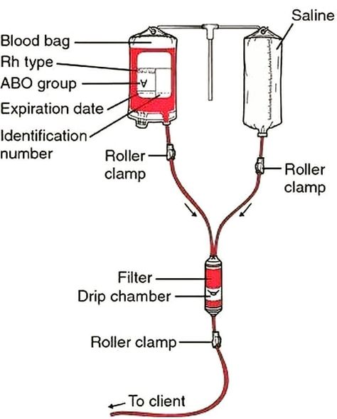
On the other hand, blood transfusion is used to replication oxygen carrying capacity and intro vascular volume, especially in patients that suffered from acute blood loss. In cardiology, this is especially relevant, particularly in acute coronary syndromes or decompensated heart failure. Doctors who actually perform blood transfusion usually prefer a more restrictive approach. In stable patients, transfusion is only considered when the patients hemoglobins level decrease lower than 7 g/dL. The main goal is to improve oxygen delivery and prevent potential adverse effects.
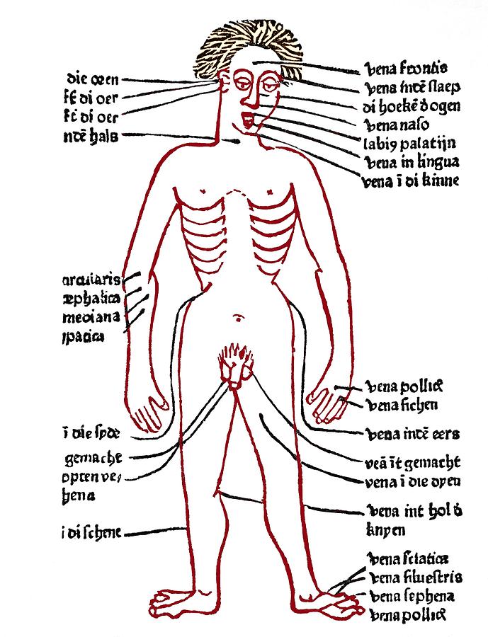
Now last but not least, bloodletting was first based on homunal theory and was before widely used across nearly every medical condition, while now it’s generally not a common treatment, and it’s more recent counterpart (therapeutic phlebotomy) remains quite relevant, particularly in hematologic disorders. In certain specific conditions such as “polycythemia Vera” and “hemochromarasis”, phlebotomy actually removes iron burden and blood viscosity. From a cardiovascular perspective, the lowering of a hematocrit helps improve micro vascular flow and reduce the risk of thrombosis as a whole, supporting circulatory efficiency. Phlebotomy is the perfect example of how older practices can be reintergrated to more modern treatments and used more effectively.
In conclusion, blood circulation, transfusion and removal are some of the most dire and necessary aspects of cardiology and cardiovascular management in general. They defined how we treat cardiology as a speciality and is the building blocks of the whole system.
Medicine has always been a huge passion for me, but it has mostly neurology and psychology that interested me the most. Yet, when I discovered cardiology, I realized just how broad and interesting all other topics of medicine are, and how useful they are, even if I won’t pursue them as a speciality.
When I was convinced me to look into cardiology topics, I was bored. I probably wasn’t going to pursue that whatsoever, so I may as well deepen my knowledge on neurological topics. Yet, when I opened the several websites and textbooks, I was transported to a world of medicine I hadn’t had a clue about before. I mean, there were simple topics from the functions of the needs of cells or the synchronization of the two pumps of the heart, to extremely complex deep dives the interventricular septum structure or the wall between ventricles or the calculations of cardiac output.
Cardiology, just like most topics in medicine, has so many sub topics for every topic there is in it, as they explore every nook and cranny of the heart.Take the venous system for example, which consists of every vein in the body, and is how veins connect with other blood vessels around the body. The venous system itself is separated into two main circuits; the systemic (left) and pulmonary (right). These both rely on blood vessels to keep blood moving and flowing through. Now in these circuits are the veins, and in the veins are the ones that carry and transport deoxygenated blood and the others that transport oxygenated blood, and this dive into veins is shallow compared to the deep dives into much more complex topics in cardiology.
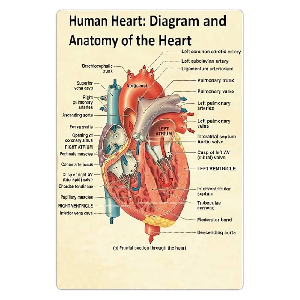
The above is one of the complex diagrams that are literally required viewing for anyone trying to get into cardiology, as the kind of diagrams simplify the relatively hard concepts, making it much easier for anyone to get into it. A simple diagram like this shows the two sides of the heart and the different arteries and sections. This also explains how blood is easily pumped and transported between the left and right ventricles, the several muscles and valves in every side and the different types of arteries, that each have a similar yet different purpose at the same time. Yet, it does get quite confusing at times due to the vast majority of cells, arteries, valves and whatnot, yet as you piece it together it really is one of the most intriguing organs in the human body.
The one thing that is amazing about this topic is the difference in people across the world. While many bodily functions function similarly or exactly the same, whereas the heart can actually significantly differ from person to person, leading to several unique conditions, such as arrhythmia (where the heartbeats are irregularly paced) or heart failure (when the heart becomes too weak to complete the conditions needed to pump blood and several other functions).
The main area of cardiology that intrigued me would easily be the electrical conduction of the heart’s system. Most organs don’t act like the heart, as the heart literally runs on its own electrical impulses, through structures such as the sinoatrrial node and atrioventricular node. Arrhythmia is actually caused by minor disruptions in this system, and it shows how fragile cardiology is. As I have said before, it’s really fascinating how something so mild can have life threatening consequences. Yet, the reason I enjoy cardiology so much is that it reaches far beyond anatomy into disease prevention. Conditions such as coronary artery disease for example, show the importantece of lifestyle management and early detection in patients, and without early detection can prove fatal. On the other hand, diseases like myocardial infractions demand immediate intervention.
This balance is what appeals to me most in this field, and the immediate impact doctors have to their patients, and how the good it does is so much more overwhelming than the bad. Cardiology can change lives, and with the research that has been developing in recent years has the opportunity to influence many more.